FACE CLINICDR AFSHIN YOUSEFPOUR
Home / The clinicFace Clinic · Digital implant dentistry

Precision by design.
The digital implant workflow.

Three-dimensional preparation for personalised treatment.

At Face Clinic in Waterloo, Dr Afshin Yousefpour combines his maxillofacial expertise with an integrated digital workflow. Imaging, impressions and planning make it possible to study your anatomy and prepare your rehabilitation before surgery.

Depending on the indication, guided surgery or dynamic navigation then supports the surgical procedure. Each tool is chosen to meet the needs of your treatment.

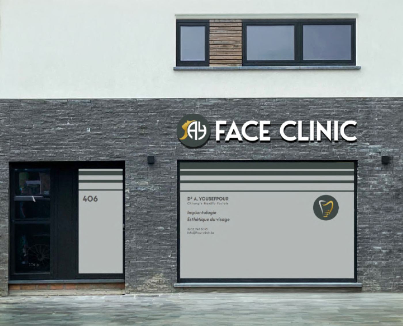
Face Clinic in Waterloo, Dr Afshin Yousefpour’s practice

NewTom GiANO HR DC: a 3D view to prepare your implants

Face Clinic is equipped with the NewTom GiANO HR DC, a 2D and 3D dental imaging system. Its CBCT (3D scan) — or cone beam — examination allows the bone structures to be studied in three dimensions to prepare a prosthetic rehabilitation on implants.

Planning implants around your future teeth

To replace one tooth, several teeth or rebuild an arch, the prosthetic plan and the anatomy must be considered together. The CBCT (3D scan) assessment helps to:

  • measure the height and width of the available bone at the planned sites;
  • locate the mandibular canal, neighbouring roots and maxillary sinuses;
  • assess the need for a bone graft or sinus lift ;
  • plan the position, angle and dimensions of the implants in relation to the future prosthesis.

This data can be combined with digital impressions and the prosthetic plan in the planning software. It is used to prepare guided or navigated surgery when indicated.

High definition and adapted acquisition

The GiANO HR platform offers several fields of view and image quality levels. The area examined and the settings are selected according to the clinical question, rather than always using the largest field or the maximum resolution.

The DC — direct conversion technology concerns the detector for 2D examinations, such as panoramic and cephalometric X-rays. It converts X-rays directly into digital signals; it is separate from 3D CBCT (3D scan) volume acquisition.

CBCT (3D scan) versus a conventional medical CT scan: what is the benefit for your treatment?

For a dental bone assessment suited to its indication, CBCT (3D scan) generally involves lower X-ray exposure than a conventional medical CT scan.

This difference is not a fixed percentage, however: it depends on the device, the volume examined, the resolution and the settings. Some medical scanners also have low-dose protocols.

Low dose: optimising exposure, keeping the useful information

The device combines several ways of controlling the dose:

  • SafeBeam™ automatically adapts the exposure to the patient’s anatomical characteristics.
  • Pulsed emission limits the X-ray emission time during 3D acquisition.
  • ECO Scan mode allows reduced-dose acquisitions when the level of detail meets the clinical need.
  • A targeted field of view limits the irradiated area to what the examination requires.

The protocol is chosen to obtain the necessary diagnostic information with optimised exposure. A detailed implant assessment may require a mode other than ECO Scan. The dose depends in particular on the field examined, the protocol and the patient: “low dose” does not mean no radiation.

Radiation protection is part of your care

At Face Clinic, the aim is to gather the information needed for implant planning while avoiding unnecessary exposure. This means justifying each examination, taking existing images into account and adapting the field and settings to your situation.

CBCT (3D scan) uses X-rays and generally involves more exposure than a 2D dental X-ray. It is used when its diagnostic value justifies it. Safety relies on this balance between clinical benefit and optimised exposure.

Radiation protection reference: FDA — Dental Cone-beam Computed Tomography.

Documentation: NewTom GiANO HR DC and manufacturer’s brochure. The available functions depend on the device’s configuration and protocols.

Recording your smile, preparing the future teeth

The digital impression records the shape of the teeth and gums with an intraoral scanner. It adds information to the bone imaging to build the restoration plan.

Digital data makes communication easier between the surgeon, the dentist making the prosthesis and the laboratory. The preparation therefore takes into account the future teeth, their position and how they fit into your smile.

OsiriX & Neowise: analysing and planning in 3D

Dr Yousefpour relies on digital analysis and planning tools, including OsiriX and Neowise, to study imaging data and prepare the implant plan.

The intended length, diameter, angle and depth of the implants are assessed according to your anatomy, the available bone and the prosthetic plan. This virtual preparation makes it possible to anticipate the stages of surgery and discuss the treatment options.

Depending on the situation, the plan can be transferred to surgery using a surgical guide or a dynamic navigation system.

X-Guide: real-time assistance

The X-Guide system can be compared to a surgical GPS: it tracks the instruments and displays their position relative to the recorded anatomy and the three-dimensional plan.

During surgery, the surgeon has information on the position, angle and depth of drilling. This interactive guidance helps him follow his plan and control the procedure as surgery progresses.

Navigation relies on rigorous registration and calibration. It assists the surgeon, who keeps control of and responsibility for the procedure at every step.

Precise preparation, attentive care

  • A plan explained before surgery. 3D visualisation helps you better understand your anatomy and the proposed choices.
  • Help with precise placement. Planning and guidance help position the implants according to the surgical and prosthetic plan.
  • An approach that respects the tissues. When the situation allows, less invasive surgery may be considered. The surgical access is still determined by your clinical needs.
  • Attention to comfort and recovery. Preparation aims to avoid unnecessary procedures and organise your follow-up. Swelling, discomfort and recovery vary with the procedure and with each patient.

Digital technology does not remove surgical risks and does not, on its own, guarantee the success or lifespan of implants. The assessment, tissue quality, hygiene and follow-up also contribute to the result.

Your implant plan

Technology makes full sense within a personalised plan.

One tooth to replace, several missing teeth or bone preparation to consider: discover the solutions, then discuss the options suited to your situation with Dr Yousefpour.

Discover your treatment options
Dental implantsDental implants in WaterlooDental implants in Braine-l’AlleudDental implants in Brussels
Face Clinic406 chaussée de Bruxelles, 1410 WaterlooWhatsApp: +32 491 07 04 32 (messages only)Google reviews
Dr Yousefpour onlinefaceexpert.be — maxillofacial surgeryInstagramLinkedIn